Warning: You are viewing this site with an outdated/unsupported browser.
Please update your browser or consider using a different one in order to view this site without issue.
For a list of browsers that this site supports, see our Supported Browsers page.
3D Video Reconstruction of Skeletal Anatomy - Robotics Institute Carnegie Mellon UniversitySkip to content
The objective of this project is to reconstruct the 3D shape of the skeleton of mouse fetuses for pharmaceutical research. The major difficulty of this project is that the fetal skeletons are stored in glass bottles filled with glycerol which causes distortion/refraction in the camera images.
Method
Since light travels in straight lines in a homogeneous medium, we can experimentally calibrate the distortion/refraction of the images caused by the glycerol and glass bottle by putting a simple calibration grid on a glass bottle filled with glycerol (but without the actual fetus bones). The grid is first imaged with it facing the camera and then imaged again with a 180 degrees rotation (so that the grid is imaged with the bottle of glycerol in between the camera and the grid to capture the distortion). Once these images are captured, the grids positions are extracted and we can estimate the directions of the light inside the bottles. Once this is known, the fetus are imaged on a rotation table and Shape-From-Silhouette is used to reconstruct the shapes of the bones.
Results
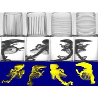
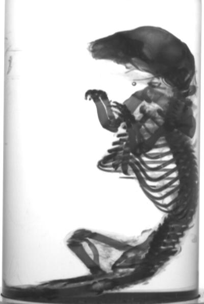
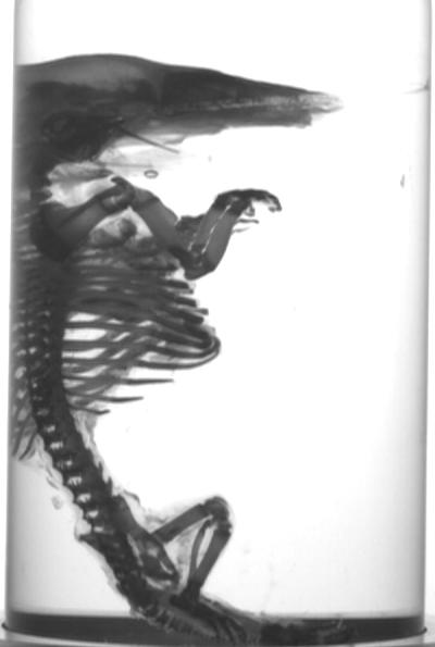
Some of the input images
The calibration grid images
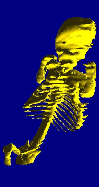
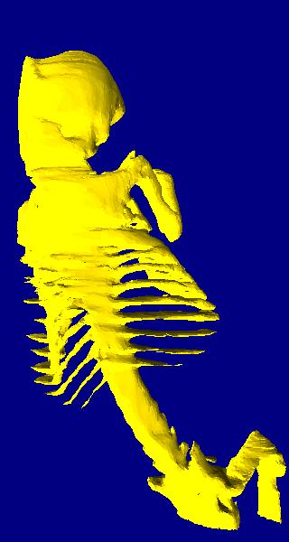
The reconstructed 3D model (from different viewpoints)