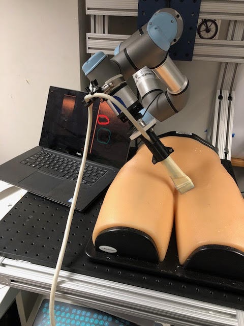
In the case of high-tempo, traumatic scenarios on the battlefield, real-time ultrasound (US) imaging serves as an enabler for countless possible robotic interventions. Having the ability to automatically segment anatomical landmarks in the body, such as arteries, veins, ligaments, and veins, for percutaneous procedures remains to be a difficult task, even in “well-controlled” settings such as hospitals where physicians and other caregivers, with considerable training, and therefore such procedures may experience complications. Performing percutaneous procedures outside of the hospital, in the field, presents additional challenges, when considering all the situations in which medical care is being delivered. These situations can be in the aftermath of a natural disaster or in a combat or hostage situation. Our goal is to create AI tools that assist the medical caregiver, either in centers of medical excellence, in rural hospitals, or in the field, deliver medical care in the form of percutaneous interventions.
We have chosen ultrasound imaging to assist, really close the loop, for needle insertion in the field because ultrasound is low-cost, portable, and radiation-free. The problem with ultrasound is that it is often difficult to interpret, therefore making percutaneous interventions even more difficult, both for human caregivers and the medical AI that supports them. Therefore, the medical AI tools that we create using ultrasound to close the loop therefore must consider the countless domains across body types, potential traumatic injury scenarios, and imaging artifacts.
For medical AI, we harness the power of deep neural networks because they can learn a more complex, non-linear function and, therefore, given sufficient training data, deep neural networks more easily discriminate among different classes. In other words, they are better function approximators. The drawback of deep neural networks is that they are data hungry, and therefore require significant amounts of diverse datasets. This can be time consuming and expensive, especially when you need a trained profession, like a physician, to label data.
This work proposes a method for enhancing deep learning models’ capabilities by generating synthetic, or augmented, data which is transformed in a manner designed to account for various body types, injury scenarios, and imaging features. Our goal is to research a learning-based data augmentation method which can adaptively generate augmented images for learning invariances across various anatomical shapes and imaging artifacts. We further explore additional perspectives with which to apply similar ideas, such as transfer learning.