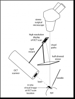
 We have developed a new image-based guidance system for microsurgery using optical coherence tomography (OCT), which presents a continuously updated virtual image in its correct location inside the scanned tissue. OCT provides real-time, 6-micron resolution images at video rates within a 2-6 mm axial range in soft or transparent tissue, and is therefore suitable for guidance to various targets in the eye. Ophthalmologic applications in general are diverse within the realm of anterior-segment surgery, whether for medical treatment or for scientific experimentation. Surgical manipulations, especially of the cornea, limbus, and lens may eventually be aided or enabled, and as an example we are presently working to guide access to Schlemm’s canal for treating Glaucoma.